
$49.99
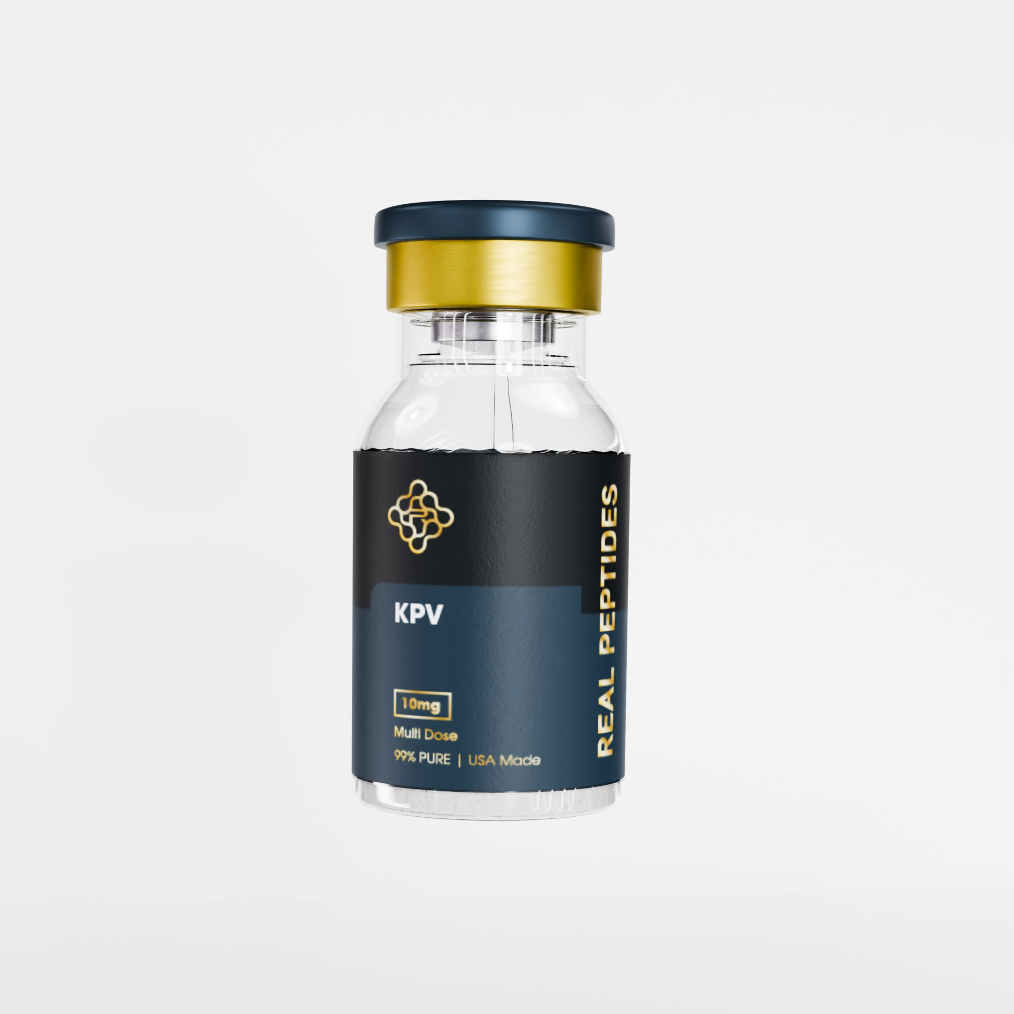
Buy KPV Peptide 10 mg Online from SourceTides. CAS 67727-97-3 | Lys-Pro-Val (H-Lys-Pro-Val-OH) | α-MSH C-terminal fragment (positions 11–13) | MW 342.43 g/mol | C₁₆H₃₀N₄O₄ | ≥99% HPLC purity | Endotoxin <1 EU/mg (LAL) | Full CoA | Lyophilised | Dry-ice cold chain. NF-κB + MAPK inhibition. PepT1-mediated intracellular delivery. Active at nanomolar concentrations. Oral efficacy in IBD colitis models (Dalmasso 2008; Gastroenterology). No melanocortin receptor activity. WADA not prohibited. For in-vitro laboratory research use only. Not for human consumption.
Buy KPV Peptide 10 mg Online | Lys-Pro-Val | α-MSH C-Terminal Fragment | ≥99% Purity | CoA | SourceTides
Buy KPV Peptide 10 mg Online from SourceTides.
KPV (Lys-Pro-Val; CAS 67727-97-3) is a synthetic tripeptide corresponding to positions 11–13 of alpha-melanocyte-stimulating hormone (α-MSH) — the C-terminal three amino acids of the parent tridecapeptide.
Despite containing just three amino acids and having a molecular weight of only 342.43 g/mol, KPV retains the powerful anti-inflammatory activity of full-length α-MSH while entirely lacking the receptor-binding sequence responsible for melanocortin effects on pigmentation, appetite, and arousal.
KPV operates through a mechanistically distinct pathway: it is transported into inflamed intestinal epithelial cells and immune cells by the PepT1 (SLC15A1) di/tripeptide transporter — a transporter that is dramatically upregulated during intestinal inflammation — and once inside, inhibits NF-κB and MAP kinase inflammatory signalling at nanomolar concentrations.
It is one of the very few research peptides for which oral bioavailability in the target tissue (inflamed colon) is actually enhanced by the disease state being studied.
Every SourceTides vial is lyophilised, tested at ≥99% HPLC purity, and ships with a full lot-specific Certificate of Analysis.
For in-vitro laboratory research use only. Not for human consumption.
KPV Peptide 10 mg — Technical Specifications
| Parameter | Specification |
|---|---|
| Common Name | KPV (Lys-Pro-Val tripeptide) |
| Full Name | Lysine-Proline-Valine; α-MSH(11-13); C-terminal α-MSH tripeptide |
| CAS Number | 67727-97-3 |
| Molecular Formula | C₁₆H₃₀N₄O₄ |
| Molecular Weight | 342.43 g/mol |
| PubChem CID | 73455 |
| Peptide Length | 3 amino acids (tripeptide); H-Lys-Pro-Val-OH; linear; all L-configured; free N- and C-terminus |
| Origin | C-terminal fragment (positions 11–13) of α-melanocyte-stimulating hormone (α-MSH; SYSMEHFRWGKPV); first identified as autonomous anti-inflammatory unit by Hiltz & Lipton (1989) |
| Key Structural Feature | Central proline residue imposes conformational rigidity; absent the His-Phe-Arg-Trp core sequence responsible for MC1R-MC4R binding — no melanocortin receptor activity at physiological concentrations |
| Primary Mechanism | NF-κB pathway inhibition; MAP kinase (MAPK/ERK/p38) suppression; PepT1-mediated intracellular delivery in inflamed epithelial and immune cells; pro-inflammatory cytokine reduction (TNF-α, IL-1β, IL-6) |
| Active Concentration (in vitro) | Active at nanomolar concentrations (10 nM shown to inhibit NF-κB in Caco2-BBE intestinal cells; Dalmasso et al. 2008) |
| PepT1 Transport | Transported into cells by PepT1 (SLC15A1); PepT1 is upregulated in inflamed colon in IBD — disease-enhanced tissue uptake; active in both intestinal epithelial cells and immune cells |
| Oral Viability | Orally active in IBD research models; tripeptide size allows PepT1-mediated intestinal uptake; oral delivery effective in DSS and TNBS colitis mouse models; gastric acid stable |
| Physical Form | White lyophilised powder; hygroscopic |
| Purity | ≥99% (RP-HPLC); identity confirmed by ESI-MS |
| Endotoxin | <1 EU/mg (LAL chromogenic assay) |
| Solubility | Freely soluble in sterile water and PBS pH 7.4; 1–5 mg/mL stock recommended; hygroscopic — keep desiccated and sealed |
| Storage — Lyophilised | −20°C long-term (stable 24 months); 2–8°C short-term (up to 4 weeks); protect from moisture; equilibrate sealed vial to room temperature before opening |
| Storage — Reconstituted | 2–8°C for up to 7 days; −20°C for longer; avoid freeze-thaw cycles; aliquot for single use |
| Certificate of Analysis | Lot-specific CoA with every order; HPLC chromatogram + MS data + endotoxin result |
| Regulatory Status | Not FDA, EMA, TGA, or Health Canada approved; research compound only; not a controlled substance in any jurisdiction |
| WADA Status | Not listed on the 2024–2025 WADA Prohibited List; not prohibited |
What Is KPV?
KPV stands for Lysine-Proline-Valine — the three C-terminal amino acids of alpha-melanocyte-stimulating hormone (α-MSH). To understand what makes KPV scientifically important, you need to understand its parent molecule and why the trimming away of ten amino acids made something more useful, not less.
α-MSH is a 13-amino acid peptide produced by the pituitary gland and locally in skin keratinocytes. It is best known for driving skin pigmentation — but it also has powerful anti-inflammatory and immunomodulatory properties. The problem with α-MSH in research and potential therapeutic contexts is that its melanocortin receptor-binding sequence (the His-Phe-Arg-Trp core at positions 6–9) is responsible not only for its anti-inflammatory effects but also for all its other biological activities: melanin synthesis, appetite regulation, sexual behaviour, and cardiovascular effects through MC1R, MC3R, MC4R, and MC5R. This broad receptor activity makes α-MSH a poor tool for studying inflammation in isolation — every experiment is confounded by multiple parallel receptor-mediated effects.
KPV resolves this problem. When researchers in the late 1980s identified that the C-terminal fragment (positions 11–13: Lys-Pro-Val) of α-MSH retained potent anti-inflammatory activity, they also observed that this minimal sequence lacked the His-Phe-Arg-Trp melanocortin receptor binding core. Hiltz and Lipton (1989) confirmed that KPV produces anti-inflammatory effects comparable to — and in some assays stronger than — full-length α-MSH, while showing no melanocortin receptor activation. The 11-13 fragment was not merely a weaker version of the parent. It was a pharmacologically cleaner version: all the anti-inflammatory signal, none of the receptor-mediated hormonal noise.
The second discovery that elevated KPV’s research significance was made decades later: KPV is a substrate for PepT1 (SLC15A1), the intestinal di/tripeptide transporter. PepT1 is expressed throughout the small intestine under normal conditions. During intestinal inflammation — Crohn’s disease, ulcerative colitis — PepT1 expression is dramatically upregulated in the colon. This means KPV is more efficiently taken up by inflamed colonic epithelial cells and immune cells than by normal tissue. The disease state enhances the therapeutic delivery. This pharmacological behaviour is mechanistically unique and explains why oral KPV is effective in IBD research models despite being a tripeptide that would normally face significant degradation in the gut. When you buy KPV Peptide 10 mg from SourceTides, you receive ≥99% HPLC-pure Lys-Pro-Val with full lot-specific CoA for in-vitro research.
KPV vs α-MSH: Why the Smaller Fragment Is the Better Research Tool
| Property | α-MSH (full-length) | KPV (C-terminal tripeptide) |
|---|---|---|
| Length | 13 amino acids | 3 amino acids |
| MW | 1,665 g/mol | 342.43 g/mol |
| Melanocortin receptor binding | Yes — MC1R, MC3R, MC4R, MC5R via His-Phe-Arg-Trp core | No — lacks the His-Phe-Arg-Trp binding pharmacophore; no MCR activity at physiological concentrations |
| Pigmentation effect (MC1R) | Yes — activates melanogenesis | None |
| Appetite / CNS effect (MC4R) | Yes — appetite suppression, arousal | None |
| Anti-inflammatory potency | Potent; active at nanomolar concentrations | Equally or more potent at NF-κB inhibition; active at nanomolar concentrations; independent of MCR |
| PepT1 transport | No — too large for di/tripeptide transporter | Yes — tripeptide size is the PepT1 substrate class; disease-enhanced uptake in inflamed colon |
| Oral efficacy in IBD models | Limited — larger peptide; systemic effects complicate GI research | Confirmed — oral KPV reduces DSS- and TNBS-induced colitis in mice (Dalmasso et al. 2008; Gastroenterology) |
| Research tool value | Multiple confounding MCR-mediated effects; difficult to attribute specific findings to anti-inflammatory mechanism | Clean anti-inflammatory probe; MCR-independent; ideal for dissecting NF-κB and MAPK pathways in isolation |
| Comparison with Melanotan-1 | Full-length α-MSH is the parent of both; Melanotan-1 targets MC1R pigmentation + NER DNA repair; KPV targets inflammation independent of MC1R | Complementary research tools from the same parent molecule with completely different applications |
How KPV Works — Mechanism of Action
Step 1 — PepT1-Mediated Cellular Entry: The Disease-Enhanced Uptake Mechanism
KPV’s entry into cells is the key to understanding its unique pharmacology. Most anti-inflammatory peptides act on cell-surface receptors — they bind from outside the cell and trigger intracellular signalling through receptor-coupled pathways. KPV works differently: it is actively transported into the cell by PepT1 (Peptide Transporter 1; SLC15A1) and then directly inhibits intracellular inflammatory signalling.
PepT1 is a proton-coupled oligopeptide transporter expressed on the apical membrane of small intestinal enterocytes. Its normal physiological function is to absorb dietary di- and tripeptides from the gut lumen — it recognises and transports any peptide of two or three amino acids, making KPV an ideal substrate. Crucially, PepT1 expression in the colon is minimal under normal conditions — but during intestinal inflammation (Crohn’s disease, ulcerative colitis), colonic PepT1 is strongly upregulated. This means KPV is preferentially taken up by inflamed tissue. The diseased colon is more permeable to KPV than healthy colon — a pharmacological property almost exactly opposite to most drugs, which face reduced bioavailability in inflamed tissue.
PepT1 is also expressed in macrophages, dendritic cells, and T-lymphocytes — and its expression in these immune cells is upregulated during inflammation. This means KPV is not only taken up by inflamed epithelial cells lining the gut, but also by the infiltrating immune cells driving the inflammatory response. This dual epithelial + immune cell uptake explains the breadth of KPV’s anti-inflammatory effects in IBD models: it reaches both the tissue barrier cells and the immune effectors causing the damage.
Step 2 — NF-κB Pathway Inhibition
Once inside the cell, KPV inhibits NF-κB (Nuclear Factor kappa-light-chain-enhancer of activated B cells) — the master transcriptional regulator of inflammatory gene expression. NF-κB controls the transcription of over 150 genes involved in inflammation, including TNF-α, IL-1β, IL-6, IL-8, COX-2, iNOS, and ICAM-1. In the resting state, NF-κB is held inactive in the cytoplasm by IκB (Inhibitor of kappa B) proteins. Pro-inflammatory stimuli (TNF-α, IL-1β, LPS) activate IKK (IκB kinase), which phosphorylates IκB and triggers its degradation, freeing NF-κB to translocate to the nucleus and drive inflammatory gene transcription.
KPV inhibits this pathway. In intestinal epithelial cells (Caco2-BBE and HT29-Cl.19A) and T-lymphocytes (Jurkat cells) stimulated with IL-1β, nanomolar concentrations of KPV reduced NF-κB activation as measured by a luciferase reporter assay — confirming intracellular NF-κB transcriptional inhibition, not just upstream receptor blockade. The landmark study (Dalmasso et al. 2008; Gastroenterology; PMC2431115) showed that 10 nM KPV significantly suppressed NF-κB activation and subsequent cytokine production (IL-8, TNF-α) in multiple human cell lines at concentrations achievable via PepT1-mediated uptake.
Step 3 — MAP Kinase (MAPK/ERK/p38) Pathway Suppression
Parallel to its NF-κB inhibition, KPV also suppresses the MAP kinase inflammatory cascade — specifically the ERK1/2 (extracellular signal-regulated kinase) and p38 MAPK pathways. Both of these kinase cascades are activated by inflammatory stimuli and feed into the same pro-inflammatory cytokine production that NF-κB drives, but through different transcription factor targets (AP-1, ATF2). By inhibiting both NF-κB and MAPK simultaneously, KPV produces a broader and more complete suppression of inflammatory gene expression than compounds that target only one pathway.
A 2025 study published in Toxicology confirmed that KPV inhibits the ERK/p38 MAPK pathway and caspase-1 activation in human keratinocytes (HaCaT cells) exposed to particulate matter (PM10) — extending the MAPK inhibition mechanism beyond intestinal research into skin and respiratory inflammation biology. This study showed KPV restored cell viability, reduced IL-1β secretion, and blocked ROS-driven MAPK activation at 50 µg/mL, demonstrating that the dual NF-κB and MAPK inhibition mechanism operates across multiple cell types and inflammatory stimuli.
Step 4 — Pro-Inflammatory Cytokine Suppression
The downstream consequence of NF-κB and MAPK inhibition is reduced production of all three primary inflammatory cytokines: TNF-α (the master cytokine of acute inflammation and IBD pathogenesis), IL-1β (the pyroptosis-initiating cytokine with roles in gut and skin inflammation), and IL-6 (the chronic inflammation mediator linking gut inflammation to systemic metabolic effects). KPV reduces the mRNA transcription and secreted protein levels of all three in multiple published cell models. In the DSS and TNBS colitis mouse studies (Dalmasso 2008; Kannengiesser 2008), oral KPV produced measurable reductions in colonic TNF-α, IL-1β, IL-6, and IL-12 at the tissue level — confirming that the in-vitro cytokine suppression translates to in-vivo anti-inflammatory tissue effects.
Step 5 — Colitis-Associated Cancer Protection (Colonic Tumorigenesis Prevention)
The most striking research finding for KPV beyond simple anti-inflammatory activity is its effect on colitis-associated cancer (CAC). IBD patients with long-standing colitis have a significantly elevated risk of colorectal cancer. The PepT1-KPV axis appears to play a role in preventing the inflammatory microenvironment that promotes colonic tumour development. Dalmasso et al. (2016; Cell Mol Gastroenterol Hepatol; PMC4957955) demonstrated that KPV dramatically reduced colonic tumorigenesis in an AOM/DSS mouse model of colitis-associated cancer — reducing tumour numbers, sizes, and overall burden. Critically, the anti-tumorigenic effect was completely abolished in PepT1-knockout mice, confirming that PepT1-mediated KPV uptake is essential for the cancer-protective effect. This finding places KPV at the intersection of GI inflammation research and colitis-associated cancer biology.
KPV Research Evidence
| Research Area | Evidence Level | Key Finding | Source |
|---|---|---|---|
| Intestinal Inflammation — PepT1 Mechanism | In vitro (Caco2-BBE, HT29, Jurkat cells) + in vivo (DSS and TNBS colitis mice; oral KPV) | 10 nM KPV inhibits NF-κB and MAPK in intestinal epithelial and immune cells; PepT1-mediated uptake confirmed; oral KPV reduces colitis severity (body weight loss, MPO, histology, cytokine mRNA) in two independent mouse colitis models | Dalmasso et al. 2008 — Gastroenterology — PMC2431115 |
| IBD Models — Melanocortin-Derived Anti-inflammatory | In vivo (TNBS and DSS colitis mice; i.p. and intracolonic KPV) | KPV reduces colonic inflammation in two standard IBD mouse models; reduced colonic damage scores, MPO activity, and IL-1β, IL-6, TNF-α levels; confirms anti-inflammatory effect via multiple routes | Kannengiesser et al. 2008 — Inflamm Bowel Dis — PubMed PMID: 18215434 |
| Colitis-Associated Cancer Prevention | In vivo (AOM/DSS murine colitis-associated cancer model; PepT1-KO vs WT mice) | KPV dramatically reduced colonic tumour number, size, and burden; effect abolished in PepT1-KO mice — confirms PepT1 dependence; identifies KPV as protective against IBD-associated carcinogenesis; PepT1 as therapeutic target for CAC | Dalmasso et al. 2016 — Cell Mol Gastroenterol Hepatol — PMC4957955 |
| Nanoparticle-Enhanced Delivery (Proof-of-Concept) | In vitro + in vivo (nanoparticle-encapsulated KPV in colitis models) | KPV in nanoparticles (1 pmol/L) more effective than free KPV (100 µmol/L in drinking water) — ~100-million-fold dose reduction; hyaluronic acid-functionalized nanoparticles (272 nm) targeted inflamed colonic cells via CD44; demonstrates targeted delivery feasibility | Laroui et al. 2010 — Gastroenterology — PubMed PMID: 20600962 |
| Skin / Keratinocyte Anti-inflammatory (2025) | In vitro (HaCaT human keratinocytes; PM10 exposure model) | KPV (50 µg/mL) restored cell viability, reduced IL-1β secretion, and inhibited ROS-driven ERK/p38 MAPK activation and caspase-1-driven pyroptosis in keratinocytes exposed to particulate matter; MAPK inhibition mechanism confirmed in skin cells | Zhang et al. 2025 — Toxicology — PubMed |
| KPV + FK506 Nanoparticle Combination (2024) | In vivo (acute and chronic colitis models; combined KPV + tacrolimus nanoparticle) | Combined KPV + FK506 in PepT1-targeted nanoparticles improved body weight, colon length, and disease activity index while reducing TNF-α, IL-1β, and IL-6 in both acute and chronic colitis models; demonstrates KPV as a combinable anti-inflammatory agent | Zhang et al. 2024 — Front Pharmacol — PubMed |
| Bronchial Epithelial Anti-inflammatory | In vitro (human bronchial epithelial cells; CALU-3 and 16HBE) | KPV inhibits cellular and systemic inflammation cues in bronchial epithelial cells; mechanism involves MC3R agonism in addition to NF-κB inhibition in this specific cell type; extends KPV anti-inflammatory research beyond GI into respiratory biology | Land SC 2012 — Int J Physiol Pathophysiol Pharmacol — PMC2095288 |
| α-MSH Fragment Anti-inflammatory Discovery | Foundational in vitro and in vivo (Hiltz & Lipton 1989 + Catania et al. review) | Original identification of KPV as autonomous anti-inflammatory unit; KPV exerts similar or stronger anti-inflammatory activity than full α-MSH without MCR activation; first demonstration of MCR-independent melanocortin anti-inflammatory mechanism | Catania et al. 2007 — PMC2095288 |
The PepT1 System: Why KPV Is Unique in IBD Research
Most anti-inflammatory drugs face a pharmacological paradox in IBD: the inflamed gut is harder to reach. Mucosal barrier disruption, altered blood flow, increased luminal degradation, and immune infiltration all reduce drug bioavailability in the target tissue. KPV navigates this paradox in reverse — PepT1 upregulation in inflamed colon means more KPV is taken up by diseased tissue than healthy tissue. The disease itself enhances delivery to the target.
How PepT1 Expression Changes in IBD
Under normal conditions, PepT1 is expressed predominantly in the small intestine — its natural role is absorbing dietary peptides. The colon has minimal PepT1 expression. In IBD patients (both Crohn’s disease and ulcerative colitis), colonic PepT1 expression is strongly upregulated in both epithelial cells and infiltrating immune cells (macrophages, T-cells). This is not a subtle change — colonic PepT1 mRNA and protein are dramatically elevated in inflamed versus non-inflamed segments of the same IBD patient. Non-inflamed colon in IBD patients has near-normal PepT1 levels; the inflamed segments show the elevated expression.
This tissue-specific upregulation creates a natural targeting mechanism for KPV: the peptide selectively accumulates in the tissue compartments with the highest inflammatory burden. When Dalmasso et al. performed PepT1-knockout mouse experiments, oral KPV completely lost its anti-colitis efficacy — confirming that PepT1 transport is not merely one route of entry but the essential mechanism. Without PepT1, KPV cannot enter the cells where NF-κB inhibition needs to occur.
Implications for Research Design
The PepT1 dependence of KPV’s activity has important implications for experimental design. For maximum anti-inflammatory signal in intestinal research models: DSS or TNBS colitis induction (which upregulates colonic PepT1) will produce stronger KPV responses than non-inflamed intestinal cell culture. Oral delivery in colitis models will produce more relevant results than systemic injection for gut-targeted research — because oral KPV reaches the inflamed colon directly via luminal PepT1 uptake. For epithelial cell culture studies: Caco2-BBE cells (which express functional PepT1) are the validated cell line for KPV mechanistic studies; non-PepT1-expressing cell lines will require higher concentrations. For confirming PepT1 dependence: PepT1-knockout cells or PepT1 siRNA knockdown provides the definitive control — as demonstrated in the CAC study where the anti-tumorigenic effect was PepT1-dependent.
What Is KPV Used for in Research?
| Research Field | Application | Why KPV |
|---|---|---|
| Inflammatory Bowel Disease (IBD) | DSS/TNBS colitis models; Crohn’s and UC cell models; mucosal healing; colon barrier function; cytokine profiling | Most extensively published anti-inflammatory peptide for intestinal research; two validated mouse colitis models; PepT1-mediated targeted delivery to inflamed colon; nanomolar active concentration; oral efficacy confirmed in vivo |
| NF-κB Biology | NF-κB pathway inhibition; IκB phosphorylation; cytokine gene transcription; NF-κB luciferase reporter assays | Active at 10 nM (extremely potent NF-κB inhibitor); MCR-independent mechanism allows clean pathway attribution; validated in multiple cell types; ideal probe for dissecting NF-κB from parallel inflammatory pathways |
| Colitis-Associated Cancer | AOM/DSS murine CAC models; colorectal carcinogenesis; tumour prevention; PepT1 oncology biology | Published RCT-level preclinical data showing KPV reduces CAC tumorigenesis (Dalmasso 2016); PepT1-dependent mechanism; connects IBD inflammation research to cancer biology; unique research compound at this mechanistic intersection |
| PepT1 Transporter Pharmacology | PepT1 substrate studies; transporter kinetics; IBD-induced PepT1 upregulation; targeted drug delivery research | KPV is the most published PepT1 substrate with documented anti-inflammatory activity; ideal probe for studying PepT1 transport biology and the therapeutic implications of intestinal inflammation-induced transporter upregulation |
| Skin Inflammation / Dermatology | Contact dermatitis models; keratinocyte inflammation; wound healing; pollution-induced skin inflammation; MAPK/NF-κB in skin cells | 2025 keratinocyte data confirms ERK/p38 MAPK and caspase-1 inhibition; no pigmentation effects (unlike Melanotan-1); clean anti-inflammatory tool for skin research without MC1R confounders; complements BPC-157 for wound healing research panels |
| Nanoparticle Drug Delivery | PepT1-targeted nanoparticle design; colon-targeted delivery; dose reduction studies; encapsulation efficacy | 100-million-fold dose reduction via nanoparticle encapsulation documented (Laroui 2010); hyaluronic acid-KPV nanoparticles validated; KPV is the canonical payload for PepT1-targeted colonic nanoparticle research |
| Respiratory Inflammation | Bronchial epithelial inflammation; airway hyperresponsiveness; pulmonary cytokine biology | Published data in human bronchial epithelial cells (Land SC 2012); extends beyond GI to respiratory biology; MC3R agonism in this tissue type adds receptor dimension not seen in gut cells |
| Gut-Brain Axis Research | Gut inflammation and neuroinflammation connections; vagal afferent signalling; intestinal cytokine-brain axis | KPV’s origin in α-MSH connects gut and neuroendocrine biology; studied alongside DSIP and Selank Amidate in gut-brain axis neuroinflammation panels |
KPV Pharmacokinetics and Experimental Considerations
| Parameter | Value / Notes | Research Implication |
|---|---|---|
| Active In-Vitro Concentration | 10 nM for NF-κB inhibition in PepT1-expressing intestinal cells; 50 µg/mL (~146 µM) for MAPK inhibition in keratinocytes; variable by cell type and endpoint | Run full dose-response (1 nM – 10 µM) in your specific cell system; PepT1-expressing cells (Caco2-BBE, HT29-Cl.19A) require lower concentrations than non-expressing cells; confirm PepT1 expression status of your cell line before comparing to published data |
| Plasma Half-Life | Short — tripeptides are rapidly degraded by plasma peptidases; in-vivo effects outlast circulating peptide due to intracellular accumulation via PepT1; exact half-life not formally characterised | Measure intracellular endpoints (NF-κB reporter, cytokine mRNA, phosphorylation states) 30–120 minutes after addition; for in-vivo colitis models, endpoint measurement (histology, MPO, cytokines) at 24–72 hours reflects sustained intracellular effects |
| Routes of Administration (research) | Oral (validated in colitis models — most relevant for IBD research); SC/IP injection (for systemic anti-inflammatory endpoint studies); topical (for skin research; active in wound healing and dermatitis models); intracolonic (for direct mucosal delivery studies) | Choose oral for IBD/gut-targeted research — PepT1 transport is the primary delivery mechanism and is active via oral route; choose SC/IP for systemic inflammatory endpoint studies; choose topical for skin inflammation research |
| Oral Dose in Colitis Models | 1–1000 µg/mouse/day in drinking water or by gavage (published range); Laroui 2010 nanoparticle study achieved 1 pmol/L equivalent; free KPV requires higher concentrations due to luminal degradation | Free KPV: start at 10 µg/mouse/day via gavage; establish dose-response from 1–1000 µg/day; nanoparticle formulation dramatically reduces required dose if targeted delivery is the research question |
| Gastric Acid Stability | Reasonably stable at gastric pH 1–2; tripeptide size and Pro residue provide some conformational protection; some degradation by intestinal peptidases expected — mitigated by PepT1 uptake at intestinal epithelium | Oral route is viable for intestinal research; for maximum mucosal bioavailability, consider enteric-coated delivery or nanoparticle encapsulation in experimental designs requiring very low doses |
| Cell Line Selection | PepT1-expressing: Caco2-BBE (best validated), HT29-Cl.19A, primary human colonocytes; Immune cells: Jurkat (T cells), THP-1 (macrophages); Non-PepT1: use as controls for PepT1-dependence confirmation | Always confirm PepT1 expression in your cell line before assuming published nanomolar concentrations apply; non-PepT1-expressing cells require micromolar concentrations for equivalent effect — which is useful as an internal negative control for mechanism confirmation |
KPV Side Effects and Safety Profile
| Concern | Evidence | Protocol Note |
|---|---|---|
| No adverse effects reported in published studies | No adverse events documented in any published KPV animal study; excellent tolerability in all colitis mouse models; no toxicity signals at any dose tested in published literature | KPV is an endogenous tripeptide fragment of a naturally occurring hormone — the body already produces and processes it; metabolites (Lys, Pro, Val) are standard amino acids with no toxicity |
| No melanocortin receptor activity | Confirmed lack of MCR binding at physiological concentrations (lacks the His-Phe-Arg-Trp pharmacophore); no pigmentation, appetite, or sexual side effects — distinct from parent α-MSH and Melanotan-1 | Clean safety advantage over full-length α-MSH analogs; pigmentation and CNS confounders absent from KPV research designs |
| Hygroscopicity handling risk | Highly hygroscopic lyophilised powder; moisture absorption causes dosing inaccuracy | Equilibrate sealed vial to room temperature before opening; weigh quickly; reseal immediately; use desiccant; same protocol as other hygroscopic tripeptides (Pinealon, Thymalin) |
| Critical data gap: no human safety data | No human clinical trials completed; all evidence from in-vitro and rodent studies; formal toxicology battery not published | Treat as a preclinical research compound with a favourable animal safety profile but insufficient human characterisation; appropriate for in-vitro research and well-designed preclinical models |
| WADA not prohibited | Not on 2024–2025 WADA Prohibited List; no performance-enhancing mechanism identified | Verify WADA list annually; list updated each year |
KPV Quality Control at SourceTides
Every batch of KPV Peptide 10 mg from SourceTides passes these tests before release. The endotoxin specification is critical for KPV specifically — LPS contamination activates NF-κB, the exact pathway KPV is studied for inhibiting, making endotoxin the most important confounder for this compound’s primary research application.
| Test | Method | Specification | Why It Matters |
|---|---|---|---|
| Purity | RP-HPLC (C18; UV 220 nm) | ≥99% peak area purity | KPV synthesis produces Lys-Pro (KP dipeptide) and Pro-Val (PV dipeptide) as common by-products; these lack the full anti-inflammatory sequence and reduce specific activity if present above 1%; ≥99% confirms correct tripeptide dominates |
| Identity | ESI-MS ([M+H]⁺ = 343.44 Da) | Confirmed MW 342.43 g/mol; H-Lys-Pro-Val-OH sequence | Confirms correct tripeptide identity vs dipeptide by-products (KP at 243 Da; PV at 215 Da); also distinguishes from the related K(D)PT peptide (D-Pro substitution) |
| Endotoxin | LAL chromogenic assay | <1 EU/mg | The most critical QC parameter for KPV: LPS activates NF-κB and the MAPK cascade — both of which KPV is studied for inhibiting. Endotoxin contamination would confound every NF-κB reporter, cytokine ELISA, and inflammatory signalling assay by activating the pathway you are measuring KPV’s ability to suppress |
| Appearance | Visual inspection | White powder; no clumping, discolouration | Clumping indicates moisture absorption; discolouration indicates oxidation or contamination; both affect dosing accuracy and potency |
| Moisture | Karl Fischer titration | <5% w/w | Low moisture ensures dosing accuracy and prevents hydrolytic degradation of the Lys-Pro peptide bond (the most hydrolysis-prone bond in the sequence) |
| Cold-Chain Dispatch | Dry-ice packaging; temperature-logged | ≤−20°C throughout | Maintains lyophilised peptide integrity; prevents hygroscopic moisture absorption during transit |
| Certificate of Analysis | Lot-specific PDF | HPLC + MS + endotoxin + moisture + dates | Required for GLP research environments; endotoxin specification particularly noted for NF-κB research use |
KPV Regulatory Status
| Jurisdiction | Status | Notes |
|---|---|---|
| USA (FDA) | Not approved; not a DEA controlled substance; research compound only | KPV has no approved indication in the USA. Not scheduled under the Controlled Substances Act. Sold as a research chemical for laboratory use only. |
| Australia (TGA) | Not listed on ARTG; research compound | Not registered as a therapeutic good. Laboratory research access only. |
| United Kingdom (MHRA) | Unlicensed; not a controlled drug; research compound | No MHRA marketing authorisation. Not listed under the Misuse of Drugs Act 1971. |
| Canada (Health Canada) | Unapproved new drug; research access only | Not a CDSA controlled substance. Not authorised for therapeutic sale. |
| European Union (EMA) | No EMA marketing authorisation; research use | No authorised medicinal product in any EU member state. |
| WADA | Not listed on the 2024–2025 WADA Prohibited List; not prohibited | No performance-enhancing classification. Not a melanocortin receptor agonist. Verify WADA list annually at wada-ama.org. |
KPV vs Related Anti-inflammatory Research Peptides
| Compound | Mechanism | Primary Research Focus | Key Difference vs KPV | SourceTides |
|---|---|---|---|---|
| KPV (this product) | NF-κB + MAPK inhibition; PepT1 transport; MCR-independent | IBD; intestinal inflammation; NF-κB biology; PepT1 pharmacology; colitis-associated cancer; skin inflammation | — | Buy KPV 10 mg |
| BPC-157 | VEGFR2/NO/FAK; angiogenesis; tissue repair | GI cytoprotection; tendon/ligament; wound healing; mucosal barrier | Both have GI protective properties but via completely different mechanisms — BPC-157 via angiogenesis/NO, KPV via NF-κB/MAPK/PepT1; often studied together in gut inflammation and healing panels; BPC-157 is WADA-prohibited, KPV is not | Buy BPC-157 Capsules |
| Melanotan-1 (afamelanotide) | MC1R agonist; cAMP-CREB-MITF; NER DNA repair; melanogenesis | Photoprotection; EPP; melanogenesis; DNA repair; skin biology | Same parent molecule (α-MSH) but completely different fragment — Melanotan-1 carries the MC1R-binding His-Phe-Arg-Trp core; KPV lacks it entirely; complementary tools for dissecting melanocortin vs non-melanocortin anti-inflammatory mechanisms | Buy Melanotan-1 10 mg |
| Selank Amidate | GABAergic gene modulation; IL-6 suppression; BDNF; tuftsin-derived immunomodulation | Anxiety; neuroimmunology; neuroinflammation; IL-6 biology | Both modulate inflammatory cytokines (IL-6) but via different systems — Selank via CNS-immune axis, KPV via GI epithelial NF-κB/PepT1; studied together in neuroinflammation-gut axis research | Buy Selank Amidate 10 mg |
| Thymalin | Thymic polypeptide; T-cell differentiation; immunosenescence | Immune ageing; T-cell biology; immunosenescence | Both address immune function but at different levels — Thymalin restores T-cell production capacity, KPV directly suppresses active inflammatory signalling; complementary in IBD (where both dysregulated T-cell responses and NF-κB overactivation are pathogenic) | Buy Thymalin 10 mg |
Peer-Reviewed References
| # | Citation | Link |
|---|---|---|
| 1 | Dalmasso G et al. (2008). PepT1-Mediated Tripeptide KPV Uptake Reduces Intestinal Inflammation. Gastroenterology. 134(1):166–178. PMID: 18061177. | PMC2431115 — Gastroenterology 2008 |
| 2 | Dalmasso G et al. (2016). Critical role of PepT1 in promoting colitis-associated cancer and therapeutic benefits of KPV in a murine model. Cell Mol Gastroenterol Hepatol. 2(3):340–357. PMC4957955. | PMC4957955 — CAC / PepT1 2016 |
| 3 | Kannengiesser K et al. (2008). Melanocortin-derived tripeptide KPV has anti-inflammatory potential in murine models of inflammatory bowel disease. Inflamm Bowel Dis. 14(3):324–331. PMID: 18215434. | PubMed PMID: 18215434 |
| 4 | Laroui H et al. (2010). Nanoparticle-encapsulated KPV reduces colitis at 100 million-fold lower dose than free KPV in drinking water. Gastroenterology. 138(5):1672–1683. PMID: 20600962. | PubMed PMID: 20600962 |
| 5 | Land SC. (2012). Inhibition of cellular and systemic inflammation in bronchial epithelial cells by melanocortin-related peptides; mechanism of KPV action and a role for MC3R agonists. Int J Physiol Pathophysiol Pharmacol. 4(2):59–73. | PMC3399072 |
| 6 | Catania A et al. (2007). α-MSH related peptides: a new class of anti-inflammatory and immunomodulating drugs. Review covering KPV discovery, mechanisms, and C-terminal fragment autonomy. PMC2095288. | PMC2095288 — α-MSH Fragment Review |
| 7 | Xiao B et al. (2017). KPV loaded into hyaluronic acid-functionalized nanoparticles (~272 nm) targets inflamed colonic epithelial cells and macrophages. Molecular Therapy. 2017. | PubMed PMID: 27804996 |
| 8 | Wikipedia: KPV tripeptide. Structure, function, anti-inflammatory mechanism, skincare research applications. | Wikipedia: KPV tripeptide |
| 9 | PubChem. KPV (Lys-Pro-Val). CID 73455. CAS 67727-97-3. National Library of Medicine. | PubChem CID 73455 |
Frequently Researched Alongside KPV
These compounds are commonly studied alongside KPV in gut inflammation, immune biology, and anti-inflammatory research:
- BPC-157 Capsules — The most common research partner for KPV in gut biology; BPC-157 addresses tissue repair and angiogenesis (VEGFR2/NO pathway) while KPV addresses active inflammatory signalling (NF-κB/MAPK/PepT1 pathway); complementary mechanisms in IBD, mucosal healing, and gut barrier research
- BPC-157 Injectable Vials — Injectable format for systemic gut and tissue repair research; paired with KPV in protocols studying combined anti-inflammatory and regenerative biology
- Melanotan-1 10 mg — The MC1R-active complement to KPV from the same parent α-MSH molecule; studying KPV (MCR-independent anti-inflammation) and Melanotan-1 (MC1R-dependent photoprotection) together allows complete dissection of the α-MSH biological repertoire
- Selank Amidate 10 mg — GABAergic anxiolytic with IL-6 suppression; studied alongside KPV in neuroinflammation-gut axis research where central (Selank/IL-6) and peripheral (KPV/NF-κB) anti-inflammatory mechanisms are studied together
- Thymalin 10 mg — Thymic T-cell bioregulator; studied alongside KPV in IBD models where both T-cell dysregulation (Thymalin target) and epithelial NF-κB overactivation (KPV target) contribute to pathogenesis
- Thymosin Alpha-1 — Thymic peptide with immune modulation and antiviral properties; studied alongside KPV in combined immune regulation and gut inflammation research panels
- Pinealon 10 mg — Neuroprotective antioxidant via SOD2/GPX1; studied with KPV in gut-brain axis research combining intestinal (KPV) and CNS (Pinealon) anti-inflammatory endpoints
- DSIP Peptide 5 mg — Sleep and antioxidant neuropeptide; studied with KPV in gut-brain axis protocols where gut inflammation (KPV target) and sleep/stress (DSIP target) intersect
- Semaglutide — GLP-1R agonist; has independent gut anti-inflammatory effects via GLP-1 receptor on intestinal immune cells; studied alongside KPV in metabolic-inflammatory overlap research (NASH, obesity-associated gut inflammation)
- LIPO-C Injectable — Multi-nutrient lipotropic; hepatic fat metabolism and gut health; studied with KPV in gut-liver axis research where intestinal inflammation (KPV) and hepatic lipid accumulation (LIPO-C) are co-studied as features of MASLD
- NAD⁺ Injectable — Sirtuin substrate; SIRT1 has anti-inflammatory effects through NF-κB deacetylation and p65 activity suppression — mechanistically complementary to KPV’s direct NF-κB inhibition; studied together in inflammation-ageing intersection research
Frequently Asked Questions
You can buy KPV Peptide 10 mg (Lys-Pro-Val; CAS 67727-97-3) directly from SourceTides. Every order includes a lot-specific Certificate of Analysis with the RP-HPLC chromatogram (≥99% purity; confirms correct tripeptide vs dipeptide by-products), ESI-MS identity confirmation (MW 342.43 Da; H-Lys-Pro-Val-OH sequence), and the LAL endotoxin result (<1 EU/mg). All vials are lyophilised and dispatched on dry-ice cold chain. See the SourceTides shipping policy for dispatch details.
KPV and Melanotan-1 both derive from α-MSH (alpha-melanocyte-stimulating hormone), but they represent opposite ends of the parent molecule and have completely different biological activities.
Melanotan-1 (afamelanotide) retains the His-Phe-Arg-Trp melanocortin receptor binding core at positions 6–9 of α-MSH — the sequence responsible for MC1R activation, melanogenesis, eumelanin production, and NER-enhanced DNA repair. It is FDA-approved (as Scenesse) for EPP. It is WADA-prohibited.
KPV is positions 11–13 (the C-terminal end) — it lacks the His-Phe-Arg-Trp core entirely. It produces no melanocortin receptor activation at physiological concentrations. Instead, it works through a completely different mechanism: PepT1-mediated intracellular delivery and direct NF-κB / MAPK pathway inhibition. It is the clean anti-inflammatory tool from α-MSH biology, stripped of all pigmentation and hormonal activity. It is not WADA-prohibited.
For researchers: use Melanotan-1 when your question is about MC1R pharmacology, melanogenesis, or UV-DNA repair. Use KPV when your question is about NF-κB inflammatory signalling, gut inflammation, or PepT1-mediated delivery. Studying them together allows complete dissection of the α-MSH biological repertoire.
KPV has three properties that make it exceptionally well-suited for IBD research specifically — and each is mechanistically distinct.
First, PepT1-mediated delivery. PepT1 is the intestinal di/tripeptide transporter that is dramatically upregulated in inflamed colon in both Crohn’s disease and ulcerative colitis. KPV is a natural substrate for PepT1 — and because PepT1 is most active where inflammation is worst, KPV accumulates preferentially in the most inflamed tissue. This is drug-to-target self-targeting at the tissue level.
Second, intracellular NF-κB inhibition. Most anti-inflammatory biologics work at cell-surface receptors. KPV enters the cell via PepT1 and directly inhibits the NF-κB transcription factor that drives production of TNF-α, IL-1β, IL-6, and COX-2 — the core inflammatory genes in IBD pathogenesis. At nanomolar concentrations. That potency is extraordinary for a tripeptide.
Third, oral efficacy. The landmark Dalmasso et al. 2008 study (Gastroenterology; PMC2431115) showed that oral KPV reduced both DSS-induced and TNBS-induced colitis in mice — confirming that the PepT1 route delivers sufficient KPV to inflamed colonic tissue to produce measurable anti-inflammatory effects. This makes KPV orally viable for IBD gut research in a way that most anti-inflammatory peptides are not. SourceTides supplies KPV 10 mg for this specific research application.
For NF-κB luciferase reporter assays, cytokine ELISA (TNF-α, IL-1β, IL-6, IL-8), MAPK phosphorylation assays (ERK, p38), and cell viability studies, ≥99% HPLC purity is the minimum. Two quality issues are specific to KPV. First, dipeptide by-products (KP and PV) appear at 243 Da and 215 Da respectively — both easily detected by MS but requiring ≥99% HPLC to control below 1% of total peak area. These dipeptides may have some biological activity of their own, confounding KPV-specific interpretations if present above trace levels. Second, and most critically: endotoxin (<1 EU/mg by LAL). LPS activates NF-κB and the MAPK cascade — the exact pathways KPV inhibits. If your KPV preparation contains endotoxin, the LPS will activate NF-κB before KPV has a chance to inhibit it, and you will likely see no KPV inhibitory effect in your reporter assay — a false negative caused by endotoxin competition. Every SourceTides KPV 10 mg batch is tested to <1 EU/mg before release.
KPV can be purchased as a research compound in all major Western jurisdictions. It is not a controlled substance in any jurisdiction — it is a tripeptide fragment of a naturally occurring hormone. In the USA, it is not DEA-scheduled. In the UK, it is not controlled under the Misuse of Drugs Act 1971. In Australia, it is available as a laboratory research compound. In Canada, it is accessible for research use. KPV is not WADA-prohibited. SourceTides supplies for in-vitro laboratory research use only. See the SourceTides shipping policy for jurisdiction-specific dispatch information.
KPV has a focused but high-quality preclinical evidence base. The key published findings:
IBD — NF-κB and MAPK inhibition (Dalmasso 2008): 10 nM KPV inhibits NF-κB and MAP kinase inflammatory pathways in human intestinal epithelial cells and T-lymphocytes; PepT1 identified as transport mechanism; oral KPV reduces DSS- and TNBS-induced colitis in mice.
IBD — Colitis-associated cancer (Dalmasso 2016): KPV dramatically reduces colonic tumorigenesis in AOM/DSS cancer model; effect abolished in PepT1-knockout mice; identifies KPV as a CAC-protective agent with PepT1 as the target.
Nanoparticle delivery (Laroui 2010): KPV in nanoparticles is ~100-million-fold more effective than free KPV in drinking water — demonstrates dramatic dose reduction possible with targeted delivery.
Skin inflammation (2025): KPV inhibits ERK/p38 MAPK and caspase-1-driven pyroptosis in human keratinocytes exposed to particulate matter pollution.
Key limitation: No human clinical trials completed. All data is in-vitro and rodent models. All references on the SourceTides KPV product page.
Equilibrate the sealed vial to room temperature before opening to prevent condensation (KPV is hygroscopic). Dissolve lyophilised KPV in sterile water or PBS (pH 7.4) to a stock of 1 mg/mL (approximately 2.92 mM given MW 342.43). KPV dissolves readily in aqueous buffers — no organic cosolvent required. Filter through a 0.22 µm syringe filter before use in cell culture (the solution is already sterile from manufacturing, but filtration is standard practice). For NF-κB reporter assays and cytokine ELISAs in PepT1-expressing intestinal cells (Caco2-BBE): start with 1–100 nM working concentration; the published active concentration is 10 nM in these cells. For keratinocyte MAPK assays: 10–100 µg/mL working range (as in 2025 HaCaT study). For in-vivo colitis models (oral gavage): dissolve 1–100 µg in 200 µL sterile saline per mouse per day — this gives the range validated in published DSS/TNBS colitis protocols. Full reconstitution notes are in the CoA with every SourceTides KPV order.
KPV and BPC-157 are both studied for gut biology but through entirely different mechanisms — which is why they are frequently studied together rather than as alternatives. KPV is an anti-inflammatory agent: it suppresses NF-κB and MAPK inflammatory signalling in inflamed epithelial and immune cells via PepT1-mediated intracellular delivery. It directly reduces cytokine production and inflammatory gene transcription in the diseased gut. BPC-157 is a tissue repair agent: it promotes angiogenesis (VEGFR2/NO/FAK pathway), restores mucosal barrier integrity, accelerates wound healing, and has gastrointestinal cytoprotective effects from its gastric origin. KPV addresses the inflammatory signalling causing gut damage; BPC-157 addresses the physical repair of the damaged gut tissue. In IBD models, the ideal research combination studies both: KPV to suppress the inflammatory driving force, BPC-157 to promote mucosal healing of the damaged tissue. BPC-157 is WADA-prohibited (S0); KPV is not. SourceTides supplies both KPV 10 mg and BPC-157 capsules.
Yes — and this is one of KPV’s most distinctive research properties. Unlike most research peptides that require injection for systemic bioavailability, KPV can be administered orally and achieves therapeutic anti-inflammatory effects in the inflamed gut via PepT1-mediated uptake. The published evidence is direct: Dalmasso et al. 2008 demonstrated that orally administered KPV (in drinking water or by gavage) reduced both DSS-induced and TNBS-induced colitis in mice at the tissue level, measured by histological inflammation scores, myeloperoxidase activity, and colonic cytokine mRNA. The PepT1 transporter expressed in inflamed colonocytes takes up KPV directly from the gut lumen — bypassing the need for systemic absorption. For gut-targeted IBD research, oral administration is therefore not just convenient but pharmacologically justified as the most relevant delivery route. Note: for systemic anti-inflammatory endpoint studies (outside the GI tract), SC/IP injection provides more reliable and quantifiable systemic exposure. All SourceTides KPV 10 mg is supplied as lyophilised powder suitable for reconstitution for any of these routes.
SourceTides accepts Visa, Mastercard, American Express, cryptocurrency, and bank transfers for institutional orders. All payments go through secure, encrypted gateways. For institutional purchase orders, bulk research procurement, or custom quantities, contact the team via the SourceTides contact page. Orders are reviewed for research compliance before dispatch.
Research Use Only
All SourceTides products, including KPV Peptide 10 mg (CAS 67727-97-3; Lys-Pro-Val), are for in-vitro laboratory research use only. They are not approved by the FDA, EMA, TGA, or Health Canada. They are not for human consumption. By purchasing, the buyer confirms authorised researcher status and accepts responsibility for compliance with all applicable laws and regulations.
| Weight | 0.01 lbs |
|---|---|
| Dimensions | 5 × 5 × 5 in |
| Purity | ≥99% (HPLC-verified) |
| Strength | 10 mg |
| Form | Lyophilised Powder |
| CAS Number | 67727-97-3 |
You may also like…

Cognitive & Mood Support
Selank Amidate Peptide 10 mg Online | ≥99% Purity | CoA Included | SourceTides
Healing & Recovery Peptides
BPC-157 500 mcg Capsules Online | ≥99% Purity | CoA Included | SourceTides
Related products
Fat Loss & Metabolic Support Peptides
Kisspeptin-10 Peptide 10 mg Online | ≥99% Purity | CoA Included | SourceTides
Immune & Antimicrobial Peptides
Thymalin Peptide 10 mg Online | Thymic Bioregulator | ≥99% Purity | CoA | SourceTides

Cognitive & Mood Support
DSIP Peptide 5 mg Online | Delta Sleep-Inducing Peptide | ≥99% Purity | CoA | SourceTides
Fat Loss & Metabolic Support Peptides
AOD-9604 Peptide Online | HGH Fragment 176-191 | ≥99% Purity | CoA Included | SourceTides

Growth Hormone Modulators Peptides
Ipamorelin Peptide 10 mg Online | ≥99% Purity | CoA Included | SourceTides


Immune & Antimicrobial Peptides
Epithalon Peptide 10 mg Online | ≥99% Purity | Certificate of Analysis Included